Can Improper Centrifugation Invalidate Test Results?
Published June 9, 2026Yes, and it happens more often than most laboratories recognize.
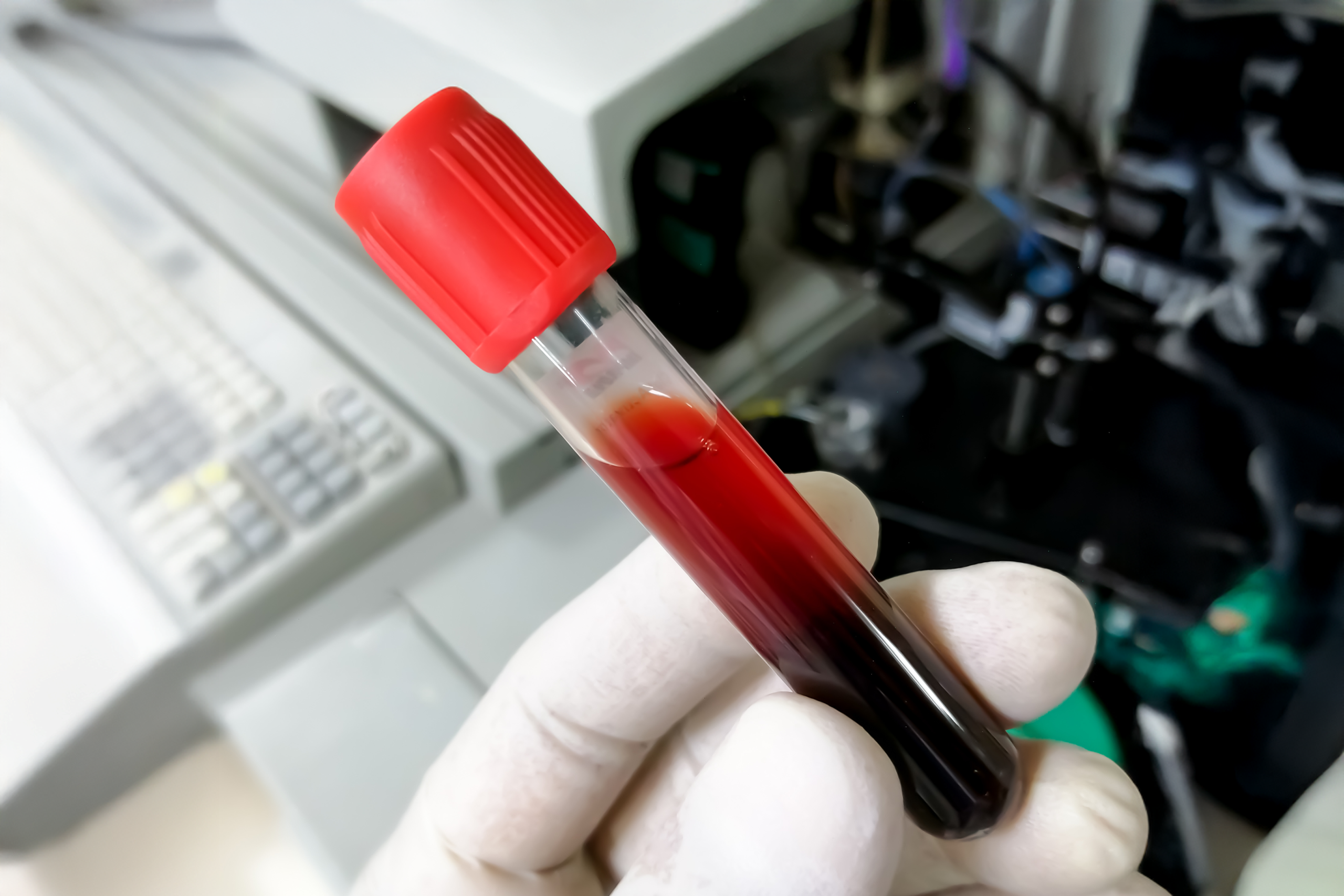
Centrifugation sits at the boundary between specimen collection and analysis, which means errors there are classified as preanalytical and are often invisible by the time a result leaves the laboratory. The sample looks processed. The analyzer ran. The number printed. But if the centrifuge step was wrong, the number may not reflect the patient at all.
TLDR
- Centrifugation errors are preanalytical, meaning they occur before the analyzer ever touches the sample.
- RCF (xg), centrifugation time, rotor type, and pre-spin clot time each independently affect result accuracy for specific analytes.
- The consequences range from a flagged hemolyzed sample to a clinically actionable false result that reaches a physician.
What does “improper centrifugation” actually mean?
Improper centrifuge is not just one thing. It is a category of errors that includes spinning too fast, too slow, too briefly, too long, at the wrong temperature, in the wrong rotor type, on equipment that is not calibrated, or without adequate pre-spin clot time. Any one of these can compromise sample integrity in ways the downstream analyzer cannot detect or correct.
Three CLSI consensus guidelines and one tube manufacturer IFU cover the parameters across application types:
- CLSI guidelines for coagulation specimens govern plasma-based coagulation testing.¹
- CLSI guidelines for chemistry specimens govern serum and plasma chemistry, including separator-tube use and analyte-by-analyte effects of hemolysis.²
- CLSI guidelines for cytology specimens govern nongynecological cytology preparation.³
- The BD Vacutainer IFU provides tube-specific RCF and time ranges.⁴
How much of the problem is preanalytical?
More than most clinicians assume. In a review of studies on laboratory errors across multiple clinical laboratory settings, % of total testing errors occur in the preanalytical phase, with .0% in the postanalytical phase and only .0% in the analytical phase itself.⁵ Preanalytical errors include sample misidentification, collection, and sample quality issues such as hemolysis, lipemia, icterus, clots, and inadequate sample volume.5
Centrifugation is a preanalytical step. That means errors occurring here are also the ones furthest from the analyzer, and the hardest to catch downstream.
What happens when you don’t spin long enough?
Under-spinning is the more common error in fast-paced clinical settings where turnaround time is under pressure. The result is incomplete separation. Cellular components remain suspended in what should be a clear plasma or serum layer.
In serum chemistry, the consequence of an under-spin is an incomplete gel barrier. CLSI guidelines for chemistry describe the gel as forming a relatively impermeable barrier between the serum and the clot during centrifugation. However, the guidelines also note that fixed-angle centrifuges form this barrier at a 45-degree may require longer spin times than in swing-bucket centrifuges.² The BD Vacutainer® Evacuated Blood Collection System IFU reinforces the same point. The presence of cells and platelets in the plasma can lead to increased variability and instability of analytes that are involved in cell-mediated metabolic processes or present at higher concentrations inside cells. Specifically, the measurement of aspartate aminotransferase, glucose, inorganic phosphorus, lactate dehydrogenase, and potassium may be affected.⁴ Without an intact barrier, these continue to drift in storage.
Premature centrifugation is its own failure mode. The BD IFU specifies minimum clotting times (60 minutes for standard serum tubes, 30 minutes for SST, 5 minutes for thrombin tubes) and notes that patients with disease-related clotting abnormalities or on anticoagulant therapy require longer.⁴ CLSI guidelines for chemistry direct that tubes be kept vertically oriented from collection until centrifugation to support complete clotting.² Spinning before clot formation is complete may leave latent fibrin in the serum that continues to form post-spin, potentially producing fibrin interferents in assays or clotting in the analyzer’s sample probe, requiring a new sample to be drawn.
The fix is following the tube manufacturer’s instructions for use (IFU) and CLSI guidance for the specific tube type and analyte. Centrifuging “until it looks done” is not a protocol.
What happens when you spin too fast or too long?
Short of that, hemolysis from the centrifuge step itself is rare. CLSI guidelines for chemistry enumerate the in vitro causes of hemolysis: difficult venipuncture, improper specimen handling (including manual clot release with a wooden applicator stick), catheter draws, and small-gauge needles attached to vacuum tubes. CLSI identifies only two recognized hemolysis pathways that result from adverse events during centrifugation: overheating in a non-refrigerated centrifuge¹ and tube breakage resulting from centrifugation above the manufacturer’s maximum RCF.⁴
measuring free hemoglobin under controlled centrifugation conditions support the same conclusion. One study directly measured free hemoglobin in the supernatant across a wide range of centrifugation parameters—forces from 90 xg to 9,300 xg, durations from 1 to 10 minutes. A single centrifugation at any of those parameters produced no significant increase in free hemoglobin compared to blood allowed to settle naturally without centrifugation.⁶ Across the entire range used in clinical practice, mechanical rupture of red cells during one properly performed spin is essentially negligible.
Where this study did see hemolysis appear in the analyzed fraction of the sample was after repeated centrifugation. Free hemoglobin in the supernatant, or plasma remained at baseline after one spin but rose significantly after three sequential spins and continued to climb through five.⁶ Even after five total centrifugations, cumulative hemolysis was approximately 0.21–0.24%, which corresponds to free hemoglobin concentrations near the lower threshold at which routine clinical chemistry analyzers begin to flag hemolysis interference. Inducing enough hemolysis to invalidate routine results through centrifugation alone would require repeated re-spinning well beyond standard practice.
Over-spinning does cause consistent problems in one area: applications with fragile cellular elements. CLSI guidelines for cytology note that cytology specimens require concentration techniques that preserve cell morphology; routine centrifugation that is fine for serum can shear cells in a cytology specimen, producing cytoplasmic vacuoles, nuclear clefting, and cellular clumping that can be misread as malignancy.³
What about coagulation testing?
Coagulation testing has the strictest centrifugation requirements in the clinical laboratory. CLSI guidelines for coagulation require platelet-poor plasma (PPP), defined as a residual platelet count below 10 × 10⁹/L. PPP is critical for lupus anticoagulant detection, heparin monitoring, and any plasma destined for freezing and re-thaw.¹
The most commonly used parameters are 1500 xg for 15 minutes at room temperature, but the standard does not mandate a single RCF/time pair. It requires only that the chosen parameters consistently produce PPP. Three primary studies cited by the standard evaluated rapid centrifugation schemes and found that shorter, higher-force spins also achieve PPP without impairing assay reliability, including one that tested 2 minutes at 4500 xg against 15 minutes at 2200 xg and found no impact on 10 critical coagulation assays.¹,⁷,⁸,⁹ The validation matters more than any specific setting.
Clot time matters for serum-based testing but not for citrate-anticoagulated coagulation specimens. Citrate prevents clotting, so coagulation tubes can be centrifuged immediately after collection. The reverse is also important: whole blood specimens for unfractionated heparin monitoring must be centrifuged within one hour of collection because of the potential for heparin neutralization by platelet factor 4, and most other coagulation specimens within four hours when stored at room temperature.¹
Under-spinning samples leave residual platelets above the PPP threshold. Transferred platelets release procoagulant material into the assay and clotting times come back falsely shortened.¹,¹⁰
Temperature is a separate failure mode. A refrigerated centrifuge inadvertently set to low temperature can cold-activate factors VII and VIII and von Willebrand factor, with documented potential to produce falsely low values mimicking hemophilia or von Willebrand disease.¹,¹¹
Rotor type affects efficiency. Swinging-bucket rotors minimize contamination of plasma with platelets and other blood cells, particularly when plasma will be frozen or removed for other testing.¹ Fixed-angle rotors are acceptable but may require extended spin speeds or duration to achieve the same separation.
The brake affects deceleration. Use of the centrifuge brake will shorten deceleration time, which could lead to slightly higher residual platelet counts, although this has been recently challenged.¹ Two primary studies cited by CLSI disagree on whether the brake meaningfully changes residual platelet counts: Daves et. al. found a small but measurable effect.¹,¹² Boissier et. al. found no impact on routine coagulation assays or platelet counts in PPP.¹,¹³ The practical implication is that brake settings should be validated locally rather than assumed transferable from another centrifuge.
Verification is required. The standard mandates that residual platelet counts be confirmed using an automated cell counter at least annually and after any change to the centrifuge.¹ Without that verification, the laboratory has no evidence its centrifuge is producing valid coagulation specimens.
The RPM vs. RCF problem
One underappreciated source of centrifugation error is the difference between RPM and RCF, and protocols that specify one when practitioners apply the other.
RPM (revolutions per minute) describes how fast the centrifuge rotor revolves. RCF (relative centrifugal force, expressed as xg) describes the actual force exerted on the sample. RCF is calculated as RCF = 1.118 × 10⁻⁵ × r × (RPM)², where r is the reference radius in millimeters.14
Because different centrifuge models have different reference radii, the same RPM setting produces different g-forces on different machines For example: a centrifuge with a 100 mm reference radius spinning at 3,000 RPM generates approximately 1,006 xg. A centrifuge with a 150 mm reference radius at the same 3,000 RPM generates approximately 1,510 xg. That is a 50% difference in force from identical RPM settings. A protocol written in RPM and followed on a centrifuge with a larger reference radius will over-spin samples; on a smaller reference radius, it will under-spin them.
CLSI and current laboratory standards recommend specifying RCF rather than RPM precisely because RCF accounts for reference radius and RPM does not.2
Can the centrifuge itself be the problem?
Yes, and CLSI requires routine verification specifically to catch this.2 The coagulation standard requires centrifuges used for coagulation specimens to be properly maintained and regularly calibrated per manufacturer recommendations.¹ Centrifuge timer accuracy should be verified using a calibrated stopwatch and centrifuge speed should be verified using a tachometer; calibration records should be documented.
Skipping these checks is how a centrifuge drifts out of spec without anyone noticing. The displayed RPM stays the same while the actual delivered force gradually changes. The errors are systematic and consistent, which means they may not trigger the outlier flags that prompt investigation of one-off mistakes.
Rotor type is independent of speed and matters separately. Horizontal (swing-out) rotors orient tubes perpendicular to the spin axis at operating speed, producing cleaner serum or plasma separation for clinical chemistry tubes because components travel the full length of the tube along the direction of applied force. The boundary between cellular and liquid layers forms perpendicular to the tube wall and stays intact on deceleration.
Fixed-angle rotors compact a pellet against the tube wall at an angle, , which can disturb the interface and resuspend cellular material into the serum or plasma layer. CLSI notes that spinning gel tubes in a fixed-angle centrifuge forms the barrier at a 45-degree angle and may not provide an adequate seal, requiring longer spin times in fixed-angle centrifuges than in swing-bucket centrifuges, and visual inspection of the tube for completeness of the barrier.²
What does proper centrifugation look like?
The short version of best practice:
- Follow the manufacturer’s protocol for each tube type. The CLSI guidelines for coagulation and chemistry both defer specific RCF and time parameters to the tube manufacturer’s IFU.¹,²
- Specify and verify RCF, not RPM. Document protocols in xg and confirm the displayed RPM corresponds to the correct RCF for the reference radius in use.
- Confirm adequate clot time before spinning serum tubes. BD specifies 60 minutes for standard serum tubes, 30 for SST, 5 for thrombin tubes, with longer times for anticoagulated patients.⁴
- Verify residual platelet count annually for coagulation specimens and after any change to the centrifuge.¹
- Use swinging-bucket rotors where practical for cleaner barrier formation and reduced cellular contamination; extend spin time if using fixed-angle.¹,²,⁴
- Verify centrifuge timer and speed with a calibrated stopwatch and tachometer on a documented schedule.¹
- Use a centrifuge designed for clinical chemistry work rather than adapting equipment from other applications.
Why this matters beyond the individual sample
Centrifugation is the critical handoff between collection and analysis. A correct draw followed by a compromised spin can negate everything that came before it, and because the error occurs upstream of the analyzer, the result that reaches the clinician may carry no flag at all.
The practical implication is that centrifuge selection and protocol design deserve the same rigor as analyzer selection. A system that removes the variables, with known spin parameters, appropriate rotor geometry, consistent RCF delivery, and reliable sample separation, reduces a substantial and often invisible source of diagnostic error.
References
- Clinical and Laboratory Standards Institute. Collection, Transport, and Processing of Blood Specimens for Testing Plasma-Based Coagulation Assays. 6th ed. CLSI guideline H21. CLSI; 2024.
- Clinical and Laboratory Standards Institute. Procedures for the Handling and Processing of Blood Specimens for Common Laboratory Tests; Approved Guideline. 4th ed. CLSI document GP44-A4. CLSI; 2010.
- Clinical and Laboratory Standards Institute. Nongynecological Cytology Specimens: Preexamination, Examination, and Postexamination Processes; Approved Guideline. 2nd ed. CLSI document GP23-A2. CLSI; 2014.
- Becton, Dickinson and Company. BD Vacutainer® Evacuated Blood Collection System Instructions for Use. BD Life Sciences — Integrated Diagnostic Solutions; Franklin Lakes, NJ.
- Plebani M. Errors in clinical laboratories or errors in laboratory medicine? Clin Chem Lab Med. 2006;44(6):750-759. doi:10.1515/CCLM.2006.123
- Mancuso JE, Jayaraman A, Ristenpart WD. Centrifugation-induced release of ATP from red blood cells. PLoS One. 2018;13(9):e0203270. doi:10.1371/journal.pone.0203270
- Wolfensberger N, Georgiou G, Giabbani E, et al. Rapid centrifugation in the routine hemostasis laboratory. Thromb Haemost. 2019;119(12):2025-2033.
- Pfaefflin A, Schuster K, Braun R. Short centrifugation to ameliorate turn-around-time in routine coagulation testing. Clin Lab. 2017;63(11):1945-1947.
- Boissier E, Sévin-Allouet M, Le Thuaut A, et al. A 2-min at 4500 g rather than a 15-min at 2200 g centrifugation does not impact the reliability of 10 critical coagulation assays. Clin Chem Lab Med. 2017;55(6):e118-e121.
- Lippi G, Rossi R, Ippolito L, et al. Influence of residual platelet count on routine coagulation, factor VIII, and factor IX testing in postfreeze-thaw samples. Semin Thromb Hemost. 2013;39(7):834-839.
- Favaloro EJ, Soltani S, McDonald J. Potential laboratory misdiagnosis of hemophilia and von Willebrand disorder owing to cold activation of blood samples for testing. Am J Clin Pathol. 2004;122(5):686-692.
- Daves M, Giacomuzzi K, Tagnin E, et al. Influence of centrifuge brake on residual platelet count and routine coagulation tests in citrated plasma. Blood Coagul Fibrinolysis. 2014;25(3):292-295.
- Boissier E, Lakhal K, Talon L, et al. The centrifuge brake impacts neither routine coagulation assays nor platelet count in platelet-poor plasma. Clin Chem Lab Med. 2020;58(9):e185-e188.
- Burtis CA, Bruns DE, Sawyer BG, eds. Tietz Fundamentals of Clinical Chemistry. 6th ed. Saunders Elsevier; 2008.