PRP, PRF, and Exosomes: Where Centrifugation Fits in the Future of Regenerative Medicine
Published June 2, 2026Regenerative medicine is moving fast, and the terminology is moving faster. In this article, we take a look at where PRP, PRF, and exosomes stand, and why the centrifuge remains the critical variable as protocols become more exacting.
TLDR
- PRP and PRF are both prepared by centrifuging a patient’s own blood; the key differences are spin speed, anticoagulant use, and the physical form of the final product.
- Exosomes are the emerging next modality, but as of 2025, no exosome product has FDA approval for injection outside of a formal clinical trial framework.
- As PRP and PRF protocols become more standardized, centrifuge performance (specifically RCF accuracy, speed consistency, and rotor geometry) directly affects concentrate quality.
What Is PRP, and How Is It Prepared?
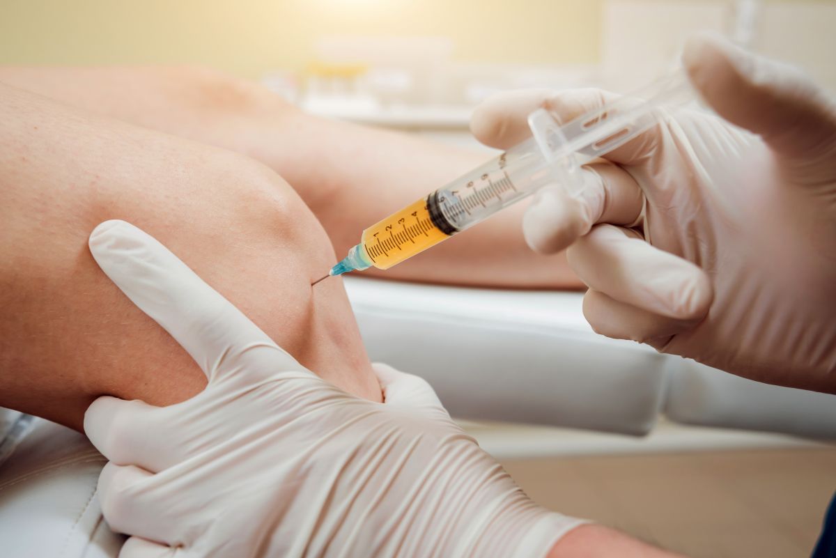
Platelet-rich plasma (PRP) is an autologous blood concentrate that delivers a higher-than-baseline concentration of platelets and growth factors to a treatment site. A small volume of blood is drawn from the patient, placed in a centrifuge, and spun to separate components by density. The platelet-rich plasma layer is extracted and prepared for injection or topical application.
The centrifuge is a critical part of the process. A 2014 review published in the Journal of Cutaneous and Aesthetic Surgery identified centrifugal acceleration, number of spins, temperature, time, and reference radius are all critical variables for optimization of PRP production and concluded each practice must standardize its own protocol.1
Because PRP uses anticoagulant-treated collection tubes, blood remains in liquid suspension throughout processing. Some PRP protocols involve two centrifugation steps: a lower-speed first spin to concentrate red blood cells to the bottom of the tube, followed by a higher-speed second spin to concentrate the platelet fraction. In one validated double-spin protocol, blood was centrifuged at The lower one-third of the final product showed maximum platelet recovery of 86–99% with a consistent 6.4 ± 0.8-fold increase over baseline.²
A 2023 randomized, double-blind trial published in the Journal of Clinical and Aesthetic Dermatology compared a single spin of 3,000 rpm for 15 minutes against a double spin of 1,500 rpm for 6 minutes followed by 2,500 rpm for 15 minutes. The single-spin method produced a 5.81 ± 1.53-fold increase in platelet concentration over baseline, significantly higher than the double-spin group with a 4.42 ± 0.94 fold increase (p = 0.012). Both methods produced high platelet yields and improved clinical outcomes, indicating that a well-specified single spin can match or exceed the platelet yield of a more complex double-spin process.¹⁷
Relative centrifugal force (RCF) is the centrifugal force produced by a centrifuge, measured in multiples of earth’s gravity.³ Because RCF is a function of both RPM and rotor radius, the same RPM setting on two different centrifuges will not produce the same g-force unless the rotor radii are identical. Three different radii can be measured on a swing-bucket rotor — minimum (top of the tube), average (center of the blood column), and maximum (bottom of the tube) —
What Is PRF, and How Does It Differ from PRP?
Platelet-rich fibrin (PRF) is a second-generation blood concentrate that originated in oral surgery before expanding into orthopedic and aesthetic applications. Two mechanical variables define it: no anticoagulant is used, and many protocols use lower centrifugation speeds.
Because blood begins clotting immediately once drawn into an anticoagulant-free tube, PRF must be processed quickly. The lower spin speed allows platelets, leukocytes, and growth factors to be captured within a fibrin matrix as it forms, rather than separated into a liquid layer. The original L-PRF protocol developed by Dr. Joseph Choukroun centrifuged blood at an RCF of 408 xg measured at the clot (RCF-max 653 xg, RCF-min 326 xg, distance to rotor for RCF-clot of 50 mm) for 12 minutes, producing a fibrin scaffold with concentrated platelets and leukocytes in the buffy coat .⁵
That fibrin structure has practical clinical advantages. Growth factors release from the matrix gradually — over days rather than the hours typical of liquid PRP — because the fibrin scaffold must degrade before bound factors are freed. Without anticoagulants, the preparation workflow is also simpler.
A study on how RCF affects PRF reports that reducing RCF from 710 xg to 177 xg led to a significantly higher number of platelets and leukocytes in the resulting concentrate. Further reduction to 44 xg showed a highly significant increase of leukocytes and platelets in comparison to both higher-speed protocols. This finding has become the basis of what researchers call the Low Speed Centrifugation Concept (LSCC) and has driven continued refinement of PRF protocols toward lower RCF values to maximize cellular content.⁶
It is worth noting that not every primary study replicates the LSCC pattern. In a head-to-head comparison of three PRF protocols on two different centrifuges, adaptation of RCF only had a minimal impact on the final characteristics of PRF membranes, and timing between blood draw and centrifugation had a larger impact than g-force adjustment.⁵ The clinically relevant point is that RCF, time, and processing delay are all material variables, not that lower RCF is uniformly superior.
Why Does Rotor Type Matter for PRF?
Rotor type is a protocol-level specification for PRF, not a minor equipment detail. In a fixed-angle centrifuge, tubes are held at a set angle (typically 33° to 45°) throughout the spin, which means cells must travel along the inclined tube wall as they separate by density. In a horizontal (swing-out) centrifuge, tubes pivot outward to a fully horizontal position during the spin, allowing blood layers to settle perpendicularly along the length of the tube.
A histological comparison of PRF clots prepared by the two systems reports that clots produced on the horizontal centrifuge showed much smoother cell layer separation along the tube surfaces compared to fixed-angle L-PRF. Horizontal centrifugation also demonstrated more evenly distributed platelets throughout the PRF clots, compared to L-PRF that gathered the majority of cells along the distal tube surface or within the buffy-coat region.7
Quantitatively, a separate study from the same group reports that horizontal centrifugation produced a significant increase in both the number and concentration of platelets and leukocytes (up to 3.5x higher for either solid or liquid PRF) when compared to either fixed-angle centrifugation systems.⁸ This finding has been cited in subsequent reviews.⁹
For practices running a standardized PRF protocol, rotor compatibility is a protocol requirement. A fixed-angle rotor will not replicate the cell-layer distribution or platelet and leukocyte yield reported for horizontal centrifugation.⁷,⁸
How Has the Push for Protocol Standardization Changed Equipment Expectations?
Early PRP and PRF adoption was marked by wide variability in preparation parameters. A systematic review of PRF centrifugation protocols across 121 published studies found 29 different centrifugation protocols and 16 different centrifuge machines in use, with reported speeds ranging from 700 to 3,500 RPM and RCF values ranging from 44 to 1,000 xg.¹² The review concluded that there was an absence of standardization in the reporting of centrifugation protocols of PRF, and proposed a reporting , AR2T3 – Angle, Radius, RCF, Time, Tube size, and Tube material – to support future meta-analyses and guidelines, which the heterogeneity of the existing data had prevented.¹⁰
This variability matters clinically because it makes outcomes from different studies nearly impossible to compare and makes it difficult for a practitioner to faithfully replicate a published protocol on different equipment. The same logic applies to PRP preparation: because the goal
The practical implication here is that a centrifuge that approximates rather than holds its programmed speed introduces the same protocol variability that standardization is designed to eliminate. A comparison of two laboratory swing-out centrifuges concluded that centrifuge type and RCF can affect the quality and quantity of cells and growth factors and an optimum relationship between g-force and RPM must be maintained in order to obtain L-PRF with adequate cell viability and optimum growth factor release.¹¹
For practitioners moving from casual PRP use to a defined, repeatable protocol, centrifuge selection has shifted from a procurement decision to a clinical one. A calibrated, reproducible g-force matters more than centrifuge brand.
What Are Exosomes, and Where Do They Stand?
The regulatory reality is a necessary counterpoint to the marketing. There are currently no FDA-approved exosome products. The FDA’s Public Safety Notification on Exosome Products directs prospective patients to ask the clinical investigator for the FDA-issued Investigational New Drug (IND) Application number and to review the FDA communication acknowledging the IND before receiving any exosome therapy.¹²
As a 2024 Clinical and Translational Science review put it, exosome therapy is in its infancy, and the industry must address safety, efficacy, and regulatory issues to realize the potential of promising exosome-based therapies. The FDA has alerted consumers that no exosome products are currently approved, and attributes ongoing market confusion to the lack of regulatory standards.¹³
One distinction practitioners often hear is that topical exosome products applied to skin post-procedure occupy a different regulatory category than injectable exosome therapies. The FDA contests this directly. As described in legal-compliance commentary on recent enforcement letters, even post-microneedling topical application is considered administration of that product and meets drug approval requirements. The only unambiguous legal pathway for therapeutic exosome use is within an FDA-authorized IND clinical trial¹⁴
Clinical investigation is ongoing. Public registries indicate that several hundred clinical trials of exosome-based therapies have been registered worldwide over the past decade and a half, though counts vary by source and inclusion criteria.¹⁵ PRP and PRF remain the more established regenerative applications.
How Does Centrifugation Connect to Exosome Research?
In research settings, centrifugation is central to exosome isolation, though at a categorically different scale. Isolating exosomes requires ultracentrifugation at 100,000 to 200,000 xg, often with density gradient steps to separate vesicle populations. This is bench-scale laboratory work, not clinical blood preparation.¹⁶
The conceptual connection is real even if the operational one isn’t: the same discipline of specifying RCF precisely — accounting for rotor radius, speed, and duration — to get reproducible output runs through PRP preparation and exosome isolation research alike. The equipment differs by orders of magnitude. The underlying principle of centrifugation as a precision instrument does not.
What Should Practitioners Know About Centrifuge Selection for PRP and PRF?
- What RCF range is needed for PRP vs. PRF? Most validated PRP protocols fall within a broad range depending on protocol design: single- or double-spin, target platelet concentration, and anticoagulant used all influence the parameters. PRF protocols generally run at lower RCF values than PRP, with the original L-PRF protocol targeting approximately 400 xg at the clot level.⁵ PRP protocols call for RCF targets up to 3,000 xg.
- Why does speed consistency matter? Because RCF is not linear with RPM, even a modest drift from the programmed speed produces a meaningfully different g-force at the sample. A centrifuge that drifts from its target speed can introduce additional variability that protocol standardization is designed to eliminate.
- Is a refrigerated centrifuge needed for PRP or PRF? Standard PRP and PRF preparation runs at room temperature. The (Association for the Advancement of Blood & Biotherapies) sets transfusion-medicine standards rather than regenerative ones, but its 21–24°C platelet-handling range is driven by platelet biology and is widely adopted for PRP for that reason.¹ While some research protocols have used refrigerated centrifuges to retard platelet activation, refrigeration adds complexity and cost without a clear consensus benefit for the production of PRP or PRF.
- What happens if the centrifuge is out of calibration? RCF is a function of both RPM and rotor radius.³ A centrifuge running even its programmed speed delivers a meaningfully different g-force to the sample — and because the relationship is nonlinear (RCF ∝ RPM²), small RPM errors produce larger RCF errors than most practitioners expect. Routine calibration verification is a basic quality control requirement in any high-volume practice.
Drucker Diagnostics manufactures centrifuges for clinical laboratory, veterinary, and regenerative medicine applications. For centrifuge specifications relevant to PRP or PRF preparation, contact our team.
References
- Dhurat R, Sukesh M. Principles and methods of preparation of platelet-rich plasma: a review and author’s perspective. J Cutan Aesthet Surg. 2014;7(4):189–197. doi:10.4103/0974-2077.150734.
- Bansal H, Leon J, Pont JL, et al. Standardization and validation of a conventional high yield platelet-rich plasma preparation protocol. Ann Med Surg. 2022;78:103834. doi:10.1016/j.amsu.2022.103834.
- Beitzel K, Russell ME. PRPCalc2: an app for platelet-rich plasma preparation. Cureus. 2023;15(2):e35193. doi:10.7759/cureus.35193.
- Dashore S, Chouhan K, Nanda S, Sharma A. Preparation of platelet-rich plasma: National IADVL PRP Taskforce recommendations. Indian Dermatol Online J. 2021;12(Suppl 1):S12–S23. doi:10.4103/idoj.idoj_269_21.
- Castro AB, Andrade C, Li X, Pinto N, Teughels W, Quirynen M. Impact of g force and timing on the characteristics of platelet-rich fibrin matrices. Sci Rep. 2021;11(1):6038. doi:10.1038/s41598-021-85736-y.
- El Bagdadi K, Kubesch A, Yu X, et al. Reduction of relative centrifugal forces increases growth factor release within solid platelet-rich-fibrin (PRF)-based matrices: a proof of concept of LSCC (low speed centrifugation concept). Eur J Trauma Emerg Surg. 2019;45(3):467–479. doi:10.1007/s00068-017-0785-7.
- Fujioka-Kobayashi M, Kono M, Katagiri H, Schaller B, Zhang Y, Sculean A, Miron RJ. Histological comparison of platelet rich fibrin clots prepared by fixed-angle versus horizontal centrifugation. Platelets. 2021;32(3):413–419. doi:10.1080/09537104.2020.1754382.
- Miron RJ, Chai J, Zheng S, Feng M, Sculean A, Zhang Y. A novel method for evaluating and quantifying cell types in platelet rich fibrin and an introduction to horizontal centrifugation. J Biomed Mater Res A. 2019;107(10):2257–2271. doi:10.1002/jbm.a.36734.
- Farshidfar N, Apaza Alccayhuaman KA, Estrin NE, Ahmad P, Sculean A, Zhang Y, Miron RJ. Advantages of horizontal centrifugation of platelet-rich fibrin in regenerative medicine and dentistry. Periodontol 2000. 2025. doi:10.1111/prd.12625.
- Herrera-Vizcaino C. Systematic review of platelet-rich fibrin (PRF) centrifugation protocols in oral and maxillofacial surgery and the introduction of AR2T3: an easy to remember acronym to correctly report vertical and horizontal PRF centrifugation. Front Oral Maxillofac Med. 2023;5:14. doi:10.21037/fomm-21-40.
- Suresh A, Balouch B, Martha VV, Sonti S. The G-force conundrum in platelet-rich fibrin generation: management of a problem hidden in plain sight. Contemp Clin Dent. 2022;13(1):3–10. doi:10.4103/ccd.ccd_697_20.
- U.S. Food and Drug Administration. Public safety notification on exosome products. December 6, 2019. Accessed via fda.gov/vaccines-blood-biologics/safety-availability-biologics/public-safety-notification-exosome-products.
- Wang CH. Regulation of exosomes as biologic medicines: regulatory challenges faced in exosome development and manufacturing processes. Clin Transl Sci. 2024;17(8):e13904. doi:10.1111/cts.13904.
- Holt DJ. Before you offer exosome therapy: understand the laws. Holt Law; October 2025. Accessed via djholtlaw.com/before-you-offer-exosome-therapy-understand-the-laws/.
- ClinicalTrials.gov. Search: “exosome” OR “extracellular vesicle.” https://clinicaltrials.gov/search?term=exosome
- Théry C, Witwer KW, Aikawa E, et al. Minimal information for studies of extracellular vesicles 2018 (MISEV2018): a position statement of the International Society for Extracellular Vesicles and update of the MISEV2014 guidelines. J Extracell Vesicles. 2018;7(1):1535750. doi:10.1080/20013078.2018.1535750.
- Legiawati L, Yusharyahya SN, Bernadette I, et al. Comparing single-spin versus double-spin platelet-rich plasma (PRP) centrifugation methods on thrombocyte count and clinical improvement of androgenetic alopecia: a preliminary, randomized, double-blind clinical trial. J Clin Aesthet Dermatol. 2023;16(12):39–44.