
Packed Cell Volume vs. Hematocrit: What They Actually Measure
Published September 30, 2026The terms packed cell volume (PCV) and hematocrit (HCT) are often used interchangeably. While the two are closely related, the methods used to obtain them can be different, which means the results from the same blood sample are not always interchangeable.
For veterinary practices running spun PCV and human laboratories using it as a backup or analyzer check, understanding how each measurement is produced can help explain differences between results and improve consistency when results are compared over time.
Packed Cell Volume and Hematocrit Are Related, but They May Not be Measured Using the Same Method
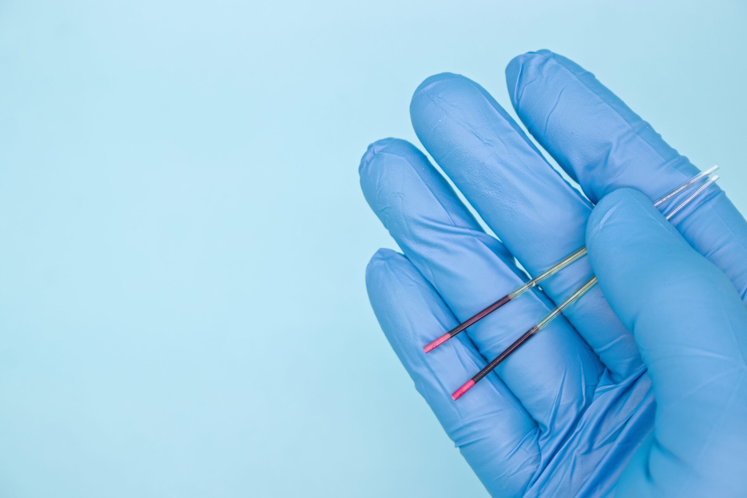
Packed cell volume is the percentage of whole blood occupied by packed red blood cells after centrifugation. With a spun microhematocrit, the PCV is read directly from the physical column of packed red blood cells in a capillary tube.
Hematocrit is the proportion of blood volume occupied by red blood cells. An automated analyzer may determine that proportion using a measurement or calculation rather than by physically packing the cells. As a result, PCV and HCT can correlate closely while still producing different numerical results.
The difference comes down to how the measurement is made. Spun PCV physically separates the blood components through centrifugation, while automated hematocrit may be calculated or measured using an analyzer-specific method.
How a Microhematocrit PCV Is Measured
A microhematocrit PCV measurement begins with a well-mixed whole-blood sample. The sample is drawn into a capillary tube, sealed, and centrifuged under validated conditions. After centrifugation, the packed red-cell column is read relative to the total blood column to determine the PCV.
When reading the packed red-cell column, measure only the packed red cells, using the top of the red-cell column, rather than the top of the buffy coat, as the boundary. The Clinical and Laboratory Standards Institute (CLSI) H07-A3 describes determination of PCV by the microhematocrit method and its use in calibration and evaluation of instrumental methods. Although the document is archived, CLSI continues to identify it as technically valid.¹
Because PCV is a physical measurement, technique can affect the result. Sample preparation, capillary filling and sealing, cell packing, and reading technique all need to be consistent.
Spin conditions can also vary by species and application. A 2025 study by Oliva et al. evaluated minimum centrifugation times for microhematocrit tubes in donkeys, dogs, sheep, and cows and found differences among the species under the study conditions. These findings should not be treated as universal settings for every veterinary application.²
How Automated Hematocrit Is Determined
“Automated HCT” does not describe one universal measurement method. Different hematology analyzers and point-of-care systems can determine hematocrit using different principles.
Some hematology analyzers calculate HCT from the measured red blood cell count and mean corpuscular volume (MCV). For example, the ADVIA 360 uses the calculation HCT = RBC × MCV / 10.³
Other analyzers measure the cumulative volume of red blood cells using electrical pulse height. Sysmex XN systems use cumulative pulse-height measurement rather than calculating HCT from RBC and MCV.⁴
Point-of-care and blood-gas systems may use another approach. The Abbott i-STAT determines HCT conductometrically from whole-blood conductivity, with a correction for electrolyte concentration.⁵
Because these methods measure or calculate HCT differently, results from different analyzers do not necessarily have the same relationship to a spun PCV.
Why PCV and Analyzer HCT Can Be Different
Even when spun PCV and analyzer HCT are measuring the same blood sample, several factors can contribute to differences between the results.
First, a small amount of plasma remains trapped within the packed red-cell column after centrifugation. Pearson and Guthrie measured a mean trapped plasma volume of 1.53% in healthy subjects, with values ranging from 1.41% to 1.82% across the patient groups studied.⁶
Technique can also affect spun PCV. Sample mixing, capillary preparation, cell packing, and reading all have an impact. A 2019 study by Breheny et al. documented variation in PCV measurements associated with inadequate sample preparation and PCV reading technique.⁷
The analyzer method itself can contribute to differences. In a 2022 comparison of four hematocrit assays, Pare et al. found that analyzer HCT methods produced lower HCT results on average than centrifuged PCV.⁸
A strong correlation between methods also does not necessarily mean the values will agree numerically. Dallarosa et al. found strong correlation between microcentrifuged PCV and conductivity-based HCT, while agreement varied by species and was poorer in dogs and cats than in horses.⁹
For that reason, a difference between a spun PCV and analyzer HCT may reflect the measurement method, sample preparation, or PCV technique rather than indicating that one result is incorrect.
Why Method Consistency Matters
PCV and analyzer HCT can have a consistent, method-dependent difference. That becomes particularly important when results are being monitored over time.
If the measurement method changes during serial monitoring, some of the apparent changes in results may come from the method rather than an actual change in the sample. When possible, keeping the measurement method consistent makes trends easier to interpret. If the method does change, that change should be identified when comparing results.
This is especially relevant when a spun PCV is used to check or compare with an analyzer result. Understanding how each result was produced provides important context when the numbers do not match exactly.
Getting a Repeatable Spun PCV
Repeatable PCV results depend on consistency throughout the process. Mixing, capillary preparation, sealing, centrifugation, and reading technique can all affect the final measurement.
A 2019 canine study found significantly less interoperator variation when staff followed a defined PCV standard operating procedure. Preparation and reading errors occurred more frequently when the SOP was not followed.⁷
Validated spin conditions should also be used for the specific sample, species, and microhematocrit system rather than assuming that one setting will work for every application.²
When selecting a centrifuge for spun PCV, confirm that it accepts the required microhematocrit tubes and can meet the validated spin conditions. RPM alone should not be used to compare centrifuges because the centrifugal force produced also depends on rotor radius.
Choosing a Centrifuge for Veterinary Microhematocrit
For practices running spun PCV regularly, or those that also process small-volume blood or urine samples, the TrueBond Duet is a great fit. Its included microhematocrit rotor holds 12 × 40 mm capillary tubes and provides a dedicated 2-minute microhematocrit cycle at 15,800 RPM and 13,700 xg.
The TrueBond Duet also includes a second rotor that accommodates two 1.5–2 mL blood or urine tubes, allowing the same centrifuge to support additional routine veterinary testing.
Drucker Diagnostics offers a risk-free 30-day evaluation program to see if TrueBond is the right fit for your practice. Try any veterinary centrifuge risk-free for 30 days, with no payment information required, before making a purchase.
Sources
- Clinical and Laboratory Standards Institute. H07-A3: Procedure for Determining Packed Cell Volume by the Microhematocrit Method; Approved Standard—Third Edition. 2000. Archived; CLSI lists the document as technically valid.
- Oliva D, Lardé H, Bouillon J, et al. “Estimation of minimum centrifugation time of microhematocrit tubes to obtain accurate results of packed cell volume and total solids in donkeys, dogs, sheep, and cows.” Journal of Veterinary Diagnostic Investigation. 2025;37(6):873–881. doi:10.1177/10406387251362461.
- Siemens Healthineers. ADVIA 360 Hematology System Operator’s Guide. HCT calculation method: RBC × MCV / 10.
- Sysmex America. XN-Series Automated Hematology Analyzers. Cumulative pulse-height hematocrit measurement. 2025.
- Abbott Point of Care. i-STAT EG6+ Cartridge Instructions for Use. Hematocrit determined conductometrically. Rev. D, September 2023.
- Pearson TC, Guthrie DL. “Trapped Plasma in the Microhematocrit.” American Journal of Clinical Pathology. 1982;78(5):770–772. doi:10.1093/ajcp/78.5.770.
- Breheny CR, Perez-Accino Salgado J, Bommer NX, Handel I, Gow AG. “Standard operating procedure reduces interoperator variation and improves accuracy when measuring packed cell volume.” Veterinary Record. 2019;184(9):283. doi:10.1136/vr.104774.
- Pare A, Kippen L, Wagg C, Longmore M, Boysen S. “Comparison of four different hematocrit assays and the effect of albumin on their measurements.” Frontiers in Veterinary Science. 2022;9:937328. doi:10.3389/fvets.2022.937328.
- Dallarosa P, Monteiro ER, Borenstein APS, Valle SF. “Agreement between hematocrit values determined by the Cobas b121 blood gas analyzer and the microhematocrit method in dogs, cats, and horses.” Veterinary Clinical Pathology. 2023;52(3):412–416. doi:10.1111/vcp.13245.