Centrifugation for PRP Preparation: Speeds, Protocols, and What Affects Platelet Yield
Published June 18, 2026The quality of a platelet-rich plasma preparation is determined long before the syringe is drawn up. Centrifuge speed, spin duration, the number of spins, and a handful of pre-analytical variables all affect how many platelets end up in the final concentrate and how biologically active they are. This guide walks through what actually drives platelet yield and what to understand when evaluating centrifuge protocols.
TLDR
- Centrifuge protocols should be specified in RCF (× g), not RPM. The same RPM produces different forces across different rotors because RCF depends on reference radius as well as speed.
- Single-spin and double-spin protocols produce different products. The data on which is better for any specific application is more contested than the popular literature suggests.
- Platelet yield is affected by more than spin parameters. Blood draw technique, anticoagulant choice, processing temperature, and the collection system all matter.
Why does the centrifuge step matter so much for PRP?
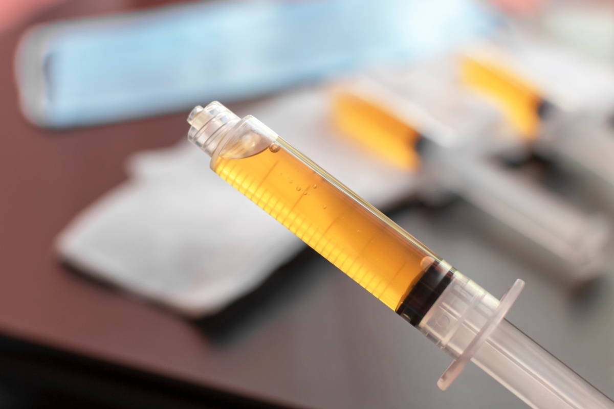
Platelet-rich plasma works by concentrating the growth factors platelets release at an injury site, including PDGF, TGF-β, VEGF, and others that drive tissue repair. The premise is straightforward: more viable, concentrated platelets mean more growth factor availability at the treatment site.
What is less straightforward is that centrifugation is where most protocols either succeed or lose consistency. A 2014 review by Dhurat and Sukesh in the Journal of Cutaneous and Aesthetic Surgery identified centrifugal acceleration, number of spins, temperature, time, and reference radius as critical variables for optimization of PRP production, and concluded each laboratory must standardize its own protocol.¹ The conclusion is worth taking seriously: protocols that work on one centrifuge configuration do not automatically transfer to another, and a substantial portion of the variability in published PRP outcomes is attributable to centrifugation differences rather than to patient biology.
For the regulatory and standardization context around this, see our separate piece on PRP protocol standardization, which covers how meta-analytic pressure and specialty society guidelines are reshaping the conversation.
RCF or RPM: which should a protocol actually specify?
RCF, every time. The distinction matters because the actual force on the sample depends on both speed and reference radius, not speed alone.
RPM (revolutions per minute) describes how fast the rotor spins. RCF (relative centrifugal force, expressed as × g) describes the force exerted on the sample. The relationship is:
RCF = 1.118 × 10⁻⁵ × r × N²
where r is the reference radius in millimeters and N is the speed in RPM.
The practical consequence is that two centrifuges running at the same displayed RPM deliver different RCF values to the sample if their reference radii differ. A protocol specifying “2,000 RPM” can therefore produce different PRP on different equipment. Drucker’s step-by-step guide to calculating G-Force walks through this in more depth. When evaluating or comparing protocols, RCF is the constant that holds across rotors; published research increasingly specifies × g for exactly this reason.\nReference radius also shapes the RCF gradient along the length of the tube, not just the average force on the sample. Because force rises with distance from the axis of rotation, the sample experiences less force at the meniscus and more at the bottom of the tube, and a smaller reference radius produces a steeper gradient across that span. A steeper gradient can make separation more sensitive to variation in hematocrit and to where the buffy coat settles along the length of the disposable, both of which can affect platelet yield.
What is the difference between single-spin and double-spin PRP?
The choice between protocols shapes the final product more than almost any other variable, but the popular framing of “double-spin is better” is more contested than it appears.
A single centrifugation separates whole blood into three visible layers: red blood cells at the bottom, the buffy coat (platelets and leukocytes) in the middle, and platelet-poor plasma at the top. The practitioner collects the buffy coat and a defined volume of the plasma above it. This produces a platelet concentrate, but the concentration factor is generally limited.
A double-spin protocol adds a second centrifugation step: the platelet-rich fraction from the first spin is transferred to a second tube and spun at higher RCF to pellet the platelets, which are then resuspended in a smaller volume of plasma. The resulting product is typically a higher-concentration platelet preparation.
The claim that double-spin reliably produces better clinical outcomes than single-spin is not as well supported as commonly assumed. A 2025 systematic review and meta-analysis in Frontiers in Medicine compared single-spin to double-spin PRP for androgenic alopecia across three randomized controlled trials and found no statistically meaningful difference in final platelet concentration between the two methods (mean difference 66.14 in single-spin compared to double-spin; 95% CI −372.96 to 505.25; p = 0.77), no meaningful difference in hair density change, and concluded that single-spin PRP may be the preferable method.² A separate 2023 randomized clinical trial in the Journal of Clinical and Aesthetic Dermatology examined thrombocyte count and clinical outcomes in single-spin versus double-spin PRP for androgenetic alopecia. Both methods raised platelet counts roughly four- to five-fold, with the larger increase in the single-spin group, and both groups showed clinical improvement in hair density and related measures.³
Researchers caution that single-spin is not universally optimal either; the appropriate protocol depends on the target application, the platelet concentration required, and the desired leukocyte composition. The takeaway is that practitioners should match the protocol to the clinical goal rather than assume the more aggressive process is the better one.
What does the buffy coat aspiration step actually achieve?
The aspiration step matters more than most protocols make clear. Even after extended centrifugation, a meaningful portion of platelets remain outside the visible buffy coat band.
Harrison and colleagues centrifuged 8.5 mL anticoagulated blood samples at 1,000 × g for spin durations from 1 to 20 minutes, then divided each sample into ten layers and counted platelets in each. They found that even after 20 minutes of centrifugation, about 15% of platelets remained in the plasma layers and about 65% in the red blood cell layers; only the remaining fraction was concentrated in the buffy coat. The highest yield came from aspirating 1 mL centered on the buffy coat (one-half mL above and one-half mL below) rather than the buffy coat alone, and that yield was reached after only 5 minutes of centrifugation.⁴
The clinical implications are practical. Spinning longer past a certain point does not necessarily move more platelets into the recovery volume, and aspirating only the visible band leaves a substantial fraction of available platelets in adjacent layers.
What about temperature and the rest of the pre-analytical chain?
Centrifuge settings are important but operate within a chain of variables. Even a well-calibrated protocol produces inconsistent results if pre-analytical technique is off.
Traumatic venipuncture activates platelets before centrifugation begins. A clean draw with minimal aspiration pressure is the starting point for any quality preparation. Pre-activated platelets degranulate during centrifugation rather than remaining intact for delivery at the treatment site.
Anticoagulant choice and ratio affect the final preparation. Most published PRP protocols use acid citrate dextrose (ACD) or sodium citrate to prevent clotting during processing. The anticoagulant-to-blood ratio matters; too little allows partial clotting and too much dilutes the preparation.
Temperature also matters. Most published PRP protocols run at room temperature. The AABB specifies a room-temperature range of 20 to 24°C for handling whole blood and platelet components, the range most PRP protocols also target.⁵ Refrigerated centrifugation can activate platelets and is generally avoided for PRP preparation. Conversely, elevated temperatures during extended processing can also compromise platelet viability.
Time from draw to processing is the last commonly overlooked variable. PRP should be centrifuged promptly after collection. Extended holding times allow platelet activation and aggregation that cannot be reversed by centrifugation.
How does the PRP protocol differ from PRF and i-PRF?
PRF (platelet-rich fibrin) is prepared differently than PRP, and the centrifuge protocol is where the difference starts. PRF uses no anticoagulant. Blood is drawn and centrifuged before natural clotting begins, allowing fibrin polymerization during the spin itself and trapping platelets and leukocytes in a fibrin matrix. The result is a solid or gel-like preparation rather than a liquid concentrate.
The original L-PRF protocol from the Choukroun group centrifuged blood at approximately 408 × g (measured at the clot) for 12 minutes.¹ For practical purposes the key differences are that PRF must be centrifuged within minutes of the draw, uses no anticoagulant, and produces a fibrin matrix rather than a liquid concentrate.
Injectable PRF (i-PRF) was developed as a liquid form. The original i-PRF protocol used a low centrifugation speed of approximately 60 × g (about 700 RPM, depending on the rotor) for 3 to 4 minutes, producing a liquid preparation that remained injectable for approximately 15 to 20 minutes before fibrin polymerization began.⁵
For a fuller comparison of PRP and PRF preparation, see our piece on PRP, PRF, and centrifugation in regenerative medicine.
What are the most common centrifugation mistakes in PRP preparation?
Three seem to recur the most across the literature and across practice.
The first is treating RPM as if it were a protocol-portable specification. RPM without reference radius context is incomplete; the same RPM on a different rotor produces different RCF. Protocols specified in × g transfer cleanly; protocols specified in RPM may not.
The second is over-centrifugation. Past the point of clean separation, spinning longer does not move more platelets into the recoverable fraction. The Harrison data is informative here: the highest platelet recovery at 1,000 × g was reached at 5 minutes, with longer durations not improving recovery materially.⁴
The third is incomplete or overly aggressive buffy coat recovery. Aspirating only the visible band leaves platelets in adjacent layers; aspirating too aggressively pulls red cells into the preparation. A defined-volume aspiration centered on the buffy coat (rather than the band alone) is the more reproducible approach.
What should a centrifuge for PRP preparation actually support?
Four capabilities matter and are worth verifying against the manufacturer’s specification rather than assumed.
The first is the ability to set RCF directly rather than calculating RPM from a published protocol. A centrifuge that accepts × g as a programmable input eliminates one source of protocol error. The G-Force calculation blog linked above covers the math when programming RCF directly is not available.
The second is gentle or no braking. Rapid braking after a spin can disturb the separation interface before aspiration begins. Centrifuges that let the rotor coast to a stop, or that offer a gentle braking profile, preserve the layer structure that aspiration depends on. Preprogrammed settings for each tube type and spin cycle extend this further, letting staff select a validated protocol rather than entering parameters by hand on every run.
The third is capacity matched to the protocol. PRP protocols vary in collection tube size, and a centrifuge that does not accommodate the tubes the protocol uses creates a workflow problem before the first sample is run.
The fourth is operational simplicity. PRP preparation is often performed by clinical staff rather than dedicated laboratory technicians. A centrifuge that ships ready to use, with the rotor and adapters configured for the relevant tube types, removes a setup-error path. Drucker’s regenerative medicine centrifuge portfolio is built around this approach, with the BOOST line designed for the range of tube configurations regenerative protocols use.
A note on regulatory context: PRP preparation protocols are typically validated against specific kits, tubes, and centrifuge configurations. Practitioners should follow the validated protocol for the system they are using rather than assume parameters from one protocol transfer to another.
Frequently asked questions
What RCF range is typical for PRP centrifugation? Published PRP protocols cover a wide range depending on protocol type, target platelet concentration, single- versus double-spin, and the desired leukocyte composition. PRP protocols vary widely, and protocol-specific parameters should be validated against the kit and tube system in use.
How long should PRP be centrifuged? Single-spin protocols generally run several minutes; double-spin protocols use a soft first spin and a harder second spin. The Harrison study found that platelet recovery at 1,000 × g reached its maximum after 5 minutes, with longer durations not improving recovery.⁴ The right duration depends on the protocol and the target product.
Does centrifuge speed affect platelet quality? Excessive force can activate or damage platelets, which reduces growth factor availability. Insufficient force leaves red blood cells in the preparation. The goal is concentrating platelets without triggering premature activation, which is why validated, protocol-specific settings matter more than general ranges.
Can the same centrifuge handle PRP and PRF protocols? A centrifuge with a sufficient RCF range and programmable settings can support both, provided the protocol-specific parameters are validated. The protocols themselves differ in speed, timing, and the absence or presence of anticoagulant, but the centrifuge requirements overlap in most respects.
For centrifuge configurations relevant to regenerative medicine applications, see our centrifuges capable of regenerative medicine work or contact the team directly.
References
- Dhurat R, Sukesh M. Principles and methods of preparation of platelet-rich plasma: a review and author’s perspective. J Cutan Aesthet Surg. 2014;7(4):189–197. doi:10.4103/0974-2077.150734.
- Ghanem L, Kirmani N, Palacios-Ortiz MP, Cevallos-Cueva M, Mendoza-Millan DL, Nascimento GVA, Velasco-Tamariz V. Comparison of single-spin to double-spin platelet-rich plasma centrifugation methods in the treatment of androgenic alopecia: a systematic review and meta-analysis of randomized controlled trials. Front Med (Lausanne). 2025. https://www.ncbi.nlm.nih.gov/pmc/articles/PMC12318733/
- Legiawati L, Yusharyahya SN, Bernadette I, Novianto E, Priyanto MH, Gliselda KC, Iriyanty S, Mutiara R. Comparing single-spin versus double-spin platelet-rich plasma (PRP) centrifugation methods on thrombocyte count and clinical improvement of androgenetic alopecia: a preliminary, randomized, double-blind clinical trial. J Clin Aesthet Dermatol. 2020. https://jcadonline.com/platelet-rich-plasma-androgenetic-alopecia/
- Harrison TE, Bowler J, Cheng CI, Reeves KD. Optimizing platelet-rich plasma: spin time and sample source. Bioengineering (Basel). 2023;10(11):1270. doi:10.3390/bioengineering10111270. https://pmc.ncbi.nlm.nih.gov/articles/PMC10669393/
- AABB. Standards for Blood Banks and Transfusion Services. 35th ed. Bethesda, MD: AABB; 2026.
- Castro AB, Andrade C, Li X, Pinto N, Teughels W, Quirynen M. Impact of g force and timing on the characteristics of platelet-rich fibrin matrices. Sci Rep. 2021;11(1):6038. doi:10.1038/s41598-021-85736-y.
- Miron RJ, Gruber R, Farshidfar N, Sculean A, Zhang Y. Ten years of injectable platelet-rich fibrin. Periodontol 2000. 2024. doi:10.1111/prd.12538.
- Marx RE. Platelet-rich plasma: evidence to support its use. J Oral Maxillofac Surg. 2004;62(4):489-496. doi:10.1016/j.joms.2003.12.003.